


PLL
La Luxation du Cristallin ou Primary Lens Luxation
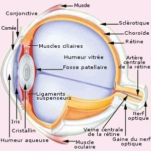
La partie transparente à l’avant du globe oculaire est appelée cornée (cornea). Autour de la cornée se situe la conjonctive (conjunctiva) et en-dessous la sclérotique (sclera). Derrière la cornée se trouve la chambre oculaire antérieure (Humeur aqueuse) remplie d’un liquide, qui sert à tenir l’oeil sous tension. Après se trouve l’iris dont la couleur varie.
Au milieu de l’iris se trouve une ouverture, c’est la pupille. Si la lumière est intense elle est contractée et petite. Quand il fait sombre elle se dilate. Le cristallin se situe derrière l’iris et est complètement encerclé par une membrane (capsule du cristallin). Quand le cristallin a une luminosité normale il est invisible à l’oeil nu et la pupille ressemble à un cercle noir au milieu de l’oeil.
Le cristallin est tenu en place par des petites fibres appelées "zonules" (Ligament suspenseurs) (Ces zonules se situent du bord du cristallin, ce bord est aussi appelé équateur, jusqu’au corps ciliaire (muscles ciliaires), ceci c’est un petit renflement qui se trouve derrière l’iris. A l’arrière du cristallin se trouve une masse gélatineuse, nommée aussi " vitré ". A l’avant de ce vitré se trouve une sorte de creux dans lequel est placé le cristallin. Ce creux est appelé " fosse patellaire ". L’oeil produit constamment de l’humidité qui est éliminée régulièrement par l’angle de filtration, qui se trouve entre la base de l’iris et la cornée. À l’arrière de l’oeil se trouvent la rétine et la pupille.
Normalement le cristallin se trouve derrière l’iris dans un aménagement du vitré. Il est tenu en place par des fibres qui se situent entre le bord du cristallin et le corps ciliaire.
La luxation du cristallin ou Primary Lens Luxation (PLL)
Elle survient lorsque le ligament fibreux de l’oeil qui maintient en place le cristallin se détériore, induisant un déplacement du cristallin de son site normal (en arrière de la pupille) vers :
- la chambre postérieure de l’œil (fig.a) : le globe oculaire se présente normalement.
- la chambre antérieure de l’œil (fig.b,c,d) : le cristallin se déplace et irrite la cornée causant un larmoiement et un écoulement bleuté dans l’œil.
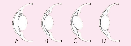
La luxation antérieure est la forme la plus hasardeuse car il existe une probabilité élevée de glaucome (augmentation de la pression du globe oculaire causée par une interruption de l’échange liquidienne - l’humeur aqueuse qui se trouve en avant du cristallin - entre le globe et la circulation veineuse. Elle mène à la cécité complète ou partielle et est de forme primaire (congénitale).
La luxation primaire du cristallin concerne des races bien déterminées à un âge bien défini. Elle attaque les deux yeux au moment où le chien ne souffre pas d’autre maladies oculaires qui pourraient être responsables du problème.
La luxation secondaire du cristallin peut se déclarer à n’importe quel âge auprès de n’importe quelle race. Une autre maladie oculaire en est responsable, peut-être le glaucome (la pression de l’oeil a d’abord augmenté et le globe oculaire s’est dilaté provoquant un basculement du cristallin). Un trauma, une grave infection oculaire, une cataracte, et des tumeurs oculaires peuvent provoquer la luxation du cristallin.
La luxation primaire (congénitale) du cristallin est autosomale récessive : chaque parent doit être au moins porteur de la maladie. Si un chien est atteint, tous ses descendants sont porteurs de la maladie.
Les chiens atteints ne doivent pas être utilisés pour la reproduction, de plus que ce désordre apparaît insidieusement entre 3-8 ans, bien au milieu du programme de l’élevage.
Les chiens porteurs sains peuvent ou pas être écartés de la reproduction, mais évitez d’accoupler deux porteurs sains ensemble : les chiots n’auraient alors que 25 % de chance d’être sains, mais 50 % seraient porteurs sains et 25 % atteints.
Cette maladie est très douloureuse pour l'animal !!!
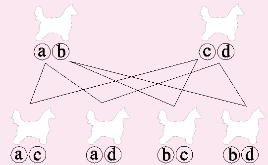
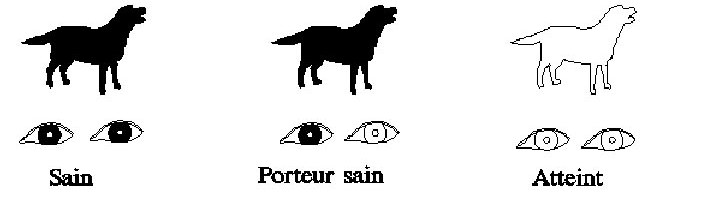
Chaque chien possède une paire de gènes susceptibles de porter des tares.
Au cours de la fécondation, chaque chien apporte un gène ; il y a donc quatre possibilités différentes :
Dominance
Le gène "sain" est dominant sur le gène "malade"
On pourra ainsi définir trois catégories de chiens :
- les chiens sains
- les chiens porteurs sains, chien porteur d’un gène "malade" mais qui ne développeront jamais la maladie.
- les chiens atteints qui développeront la maladie
Observez votre chien avant l’apparition de la luxation, vous constaterez éventuellement un changement de comportement : se cogner contre des objets, manquer une marche d’escalier, pas de poursuite de la balle ou du biscuit etc …
Le traitement est variable :
- excision du cristallin, ce qui permet une vision partielle (cher et pas toujours efficace).
- énucléation de l’œil, pour les cas sévères.
La PLL est une maladie récessive : cela veut dire que deux parents porteurs de la maladie mais qui ne déclareront jamais la PLL peuvent engendrer des chiots qui seront plus tard atteints. Voici un tableau pour vous aidez à mieux comprendre le mode de transmission:
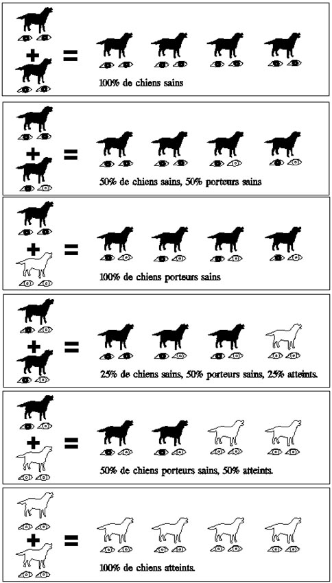
Exemple : un étalon (ou une femelle reproductrice) dont le père (ou la mère ) est luxe sera soit atteint de la luxation du cristallin (50 % de risques) ou sera porteur sain ( les 50 % restants) ; c ’ est a dire que ce chien sera prédisposé a transmettre cette maladie a sa descendance.
Une mutation qui provoque le développement de la luxation primaire du cristallin (PLL) dans de nombreuses races de chiens a été identifié par une équipe de chercheurs dirigée par les Drs Gary Johnson & Elizabeth Giuliano à l’Université du Missouri "College of Veterinary Medicine". Un test ADN pour cette mutation est devenu disponible à la mi-Septembre 2009, à travers un partenariat avec l’OFA (Orthopedic Fondation for Animals).
Peu de temps après l’annonce par l’Université du Missouri, des chercheurs de "l’Animal Health Trust en Angleterre", ont également annoncé qu’ils avaient trouvé une mutation pour la PLL. Dr Catherine Mellersh et le Dr David Sargen de l’AHT contacté le Dr Johnson, et les deux équipes de recherche ont convenu de partager les données et de co-publier cette découverte. PLL tests seront également disponibles par l’intermédiaire du AHT en Angleterre, à un prix comparable à la taxe à l’OFA.
Pour commander le test en France : https://antagene.com/ ou https://www.genimal.com/fr/ ou autres laboratoires.
Ici une copie du test des parents ou des ascendants vous sera transmis si vous le souhaitez le jour où vous venez chercher votre bébé. Sinon tout est déclaré à la SCC et donc les tests des parents ou ascendants sont notés sur le certificat de naissance de votre bébé.
Pour commencer, un peu d’anatomie de l’oeil
La partie transparente à l’avant du globe oculaire est appelée cornée (cornea). Autour de la cornée se situe la conjonctive (conjunctiva) et en-dessous la sclérotique (sclera). Derrière la cornée se trouve la chambre oculaire antérieure (Humeur aqueuse) remplie d’un liquide, qui sert à tenir l’oeil sous tension. Après se trouve l’iris dont la couleur varie.
Au milieu de l’iris se trouve une ouverture, c’est la pupille. Si la lumière est intense elle est contractée et petite. Quand il fait sombre elle se dilate. Le cristallin se situe derrière l’iris et est complètement encerclé par une membrane (capsule du cristallin). Quand le cristallin a une luminosité normale il est invisible à l’oeil nu et la pupille ressemble à un cercle noir au milieu de l’oeil.
Le cristallin est tenu en place par des petites fibres appelées "zonules" (Ligament suspenseurs) (Ces zonules se situent du bord du cristallin, ce bord est aussi appelé équateur, jusqu’au corps ciliaire (muscles ciliaires), ceci c’est un petit renflement qui se trouve derrière l’iris. A l’arrière du cristallin se trouve une masse gélatineuse, nommée aussi " vitré ". A l’avant de ce vitré se trouve une sorte de creux dans lequel est placé le cristallin. Ce creux est appelé " fosse patellaire ". L’oeil produit constamment de l’humidité qui est éliminée régulièrement par l’angle de filtration, qui se trouve entre la base de l’iris et la cornée. À l’arrière de l’oeil se trouvent la rétine et la pupille.
Normalement le cristallin se trouve derrière l’iris dans un aménagement du vitré. Il est tenu en place par des fibres qui se situent entre le bord du cristallin et le corps ciliaire.
La luxation du cristallin ou Primary Lens Luxation (PLL)
Elle survient lorsque le ligament fibreux de l’oeil qui maintient en place le cristallin se détériore, induisant un déplacement du cristallin de son site normal (en arrière de la pupille) vers :
- la chambre postérieure de l’œil (fig.a) : le globe oculaire se présente normalement.
- la chambre antérieure de l’œil (fig.b,c,d) : le cristallin se déplace et irrite la cornée causant un larmoiement et un écoulement bleuté dans l’œil.
La luxation antérieure est la forme la plus hasardeuse car il existe une probabilité élevée de glaucome (augmentation de la pression du globe oculaire causée par une interruption de l’échange liquidienne - l’humeur aqueuse qui se trouve en avant du cristallin - entre le globe et la circulation veineuse. Elle mène à la cécité complète ou partielle et est de forme primaire (congénitale).
La différence entre luxation primaire et luxation secondaire du cristallin
La luxation primaire du cristallin concerne des races bien déterminées à un âge bien défini. Elle attaque les deux yeux au moment où le chien ne souffre pas d’autre maladies oculaires qui pourraient être responsables du problème.
La luxation secondaire du cristallin peut se déclarer à n’importe quel âge auprès de n’importe quelle race. Une autre maladie oculaire en est responsable, peut-être le glaucome (la pression de l’oeil a d’abord augmenté et le globe oculaire s’est dilaté provoquant un basculement du cristallin). Un trauma, une grave infection oculaire, une cataracte, et des tumeurs oculaires peuvent provoquer la luxation du cristallin.
Une maladie autosomale récessive
La luxation primaire (congénitale) du cristallin est autosomale récessive : chaque parent doit être au moins porteur de la maladie. Si un chien est atteint, tous ses descendants sont porteurs de la maladie.
Les chiens atteints ne doivent pas être utilisés pour la reproduction, de plus que ce désordre apparaît insidieusement entre 3-8 ans, bien au milieu du programme de l’élevage.
Les chiens porteurs sains peuvent ou pas être écartés de la reproduction, mais évitez d’accoupler deux porteurs sains ensemble : les chiots n’auraient alors que 25 % de chance d’être sains, mais 50 % seraient porteurs sains et 25 % atteints.
Cette maladie est très douloureuse pour l'animal !!!
Distribution des gènes
Chaque chien possède une paire de gènes susceptibles de porter des tares.
Au cours de la fécondation, chaque chien apporte un gène ; il y a donc quatre possibilités différentes :
Dominance
Le gène "sain"
est dominant sur le gène "malade" On pourra ainsi définir trois catégories de chiens :
- les chiens sains
- les chiens porteurs sains, chien porteur d’un gène "malade" mais qui ne développeront jamais la maladie.
- les chiens atteints qui développeront la maladie
Observez votre chien avant l’apparition de la luxation, vous constaterez éventuellement un changement de comportement : se cogner contre des objets, manquer une marche d’escalier, pas de poursuite de la balle ou du biscuit etc …
Le traitement est variable :
- excision du cristallin, ce qui permet une vision partielle (cher et pas toujours efficace).
- énucléation de l’œil, pour les cas sévères.
La PLL est une maladie récessive : cela veut dire que deux parents porteurs de la maladie mais qui ne déclareront jamais la PLL peuvent engendrer des chiots qui seront plus tard atteints. Voici un tableau pour vous aidez à mieux comprendre le mode de transmission:
Exemple : un étalon (ou une femelle reproductrice) dont le père (ou la mère ) est luxe sera soit atteint de la luxation du cristallin (50 % de risques) ou sera porteur sain ( les 50 % restants) ; c ’ est a dire que ce chien sera prédisposé a transmettre cette maladie a sa descendance.
Un test ADN est maintenant disponible !!
Une mutation qui provoque le développement de la luxation primaire du cristallin (PLL) dans de nombreuses races de chiens a été identifié par une équipe de chercheurs dirigée par les Drs Gary Johnson & Elizabeth Giuliano à l’Université du Missouri "College of Veterinary Medicine". Un test ADN pour cette mutation est devenu disponible à la mi-Septembre 2009, à travers un partenariat avec l’OFA (Orthopedic Fondation for Animals).
Peu de temps après l’annonce par l’Université du Missouri, des chercheurs de "l’Animal Health Trust en Angleterre", ont également annoncé qu’ils avaient trouvé une mutation pour la PLL. Dr Catherine Mellersh et le Dr David Sargen de l’AHT contacté le Dr Johnson, et les deux équipes de recherche ont convenu de partager les données et de co-publier cette découverte. PLL tests seront également disponibles par l’intermédiaire du AHT en Angleterre, à un prix comparable à la taxe à l’OFA.
Pour commander le test en France : https://antagene.com/ ou https://www.genimal.com/fr/ ou autres laboratoires.
Ici une copie du test des parents ou des ascendants vous sera transmis si vous le souhaitez le jour où vous venez chercher votre bébé. Sinon tout est déclaré à la SCC et donc les tests des parents ou ascendants sont notés sur le certificat de naissance de votre bébé.